Trends
Other sections
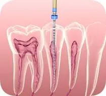
To assess the prevalence of accessory canals
and morphological variations in maxillary first molars of Saudi patients
attending private university hospitals in Riyadh using Cone-Beam Computed
Tomography (CBCT).
Methods:
This retrospective study analyzed CBCT scans of 250 patients (125 males, 125
females) aged 18-50 years, obtained from private university hospitals in Riyadh
between 2019 and 2024. Patients were divided into two age groups: 18-38 years
and 39-50 years. Root canal configurations were classified according to Weine's
classification, and isthmuses were categorized based on Hsu and Kim's
classification. Statistical analysis included descriptive statistics,
chi-square tests, and t-tests.
Results: The overall prevalence of accessory canals in
maxillary first molars was 76%. Males showed a higher prevalence (57.3%)
compared to females (42.3%), although this difference was not statistically
significant (p>0.05). No significant differences were observed between
different age groups or between right and left sides. Regarding root canal
morphology, Type I configuration (66%) was most common, followed by Type II
(32%), with Types III and IV accounting for only 1% each. For isthmus
configuration, Type IV was most prevalent (31.6%), followed by absence of
isthmus (26%), Type I (25.6%), Type II (7.2%), Type III (6.8%), and Type V
(2.8%). No statistically significant differences in root canal or isthmus
configurations were found between genders, age groups, or sides.
Conclusions:
The high prevalence of accessory canals (76%) in maxillary first molars among
Saudi patients highlights the importance of thorough examination and knowledge
of root canal morphology for successful endodontic treatment. CBCT proves to be
a valuable tool for detecting these anatomical variations. No significant
differences in prevalence or morphological variations were found between
genders, age groups, or right and left sides.